

A dental exam is a comprehensive oral health evaluation covering your teeth, gums, bite, jaw joints, and soft tissues to detect problems early and guide personalized care. Most patients think a checkup is just about cavities. In reality, what dentists check during exam appointments goes much further, including periodontal probing, oral cancer screening, TMJ assessment, and selective X-rays. Understanding each step helps you walk in prepared and walk out with a clearer picture of your oral health. At Woodbridgedentalcentre, we see patients from Woodbridge, Vaughan, and Maple who are often surprised by how thorough a well-run exam truly is.
What happens at the start of a dental exam?
Every exam begins with a review of your medical and dental history. This step shapes everything that follows. A dentist who knows you take blood thinners, have diabetes, or grind your teeth at night will approach the exam differently than one who does not. That context is not just background information. It directly affects diagnosis and treatment planning.
During this initial phase, your dentist or a team member will typically ask about:
- Any new medications or supplements you are taking
- Changes in your general health since your last visit
- Symptoms you have noticed, such as tooth sensitivity, jaw pain, or bleeding gums
- Your current brushing and flossing habits
- Any dental anxiety or concerns you want to address
Comprehensive dental exams are distinguished from periodic exams by detailed charting, complete periodontal probing, oral pathology screening, and radiographic imaging that establishes a full baseline. If you are a new patient or returning after a long absence, expect a more thorough intake process. Knowing the difference between a comprehensive and a periodic exam helps you set realistic expectations before you sit in the chair.
Pro Tip: Bring a list of your current medications to every appointment. This saves time and gives your dentist accurate information to work with from the start.
How do dentists examine teeth and gums?
The visual and tactile inspection of your teeth and gums is the core of any dental checkup. Your dentist uses a small mirror and explorer tool to check each tooth surface for signs of decay, cracks, enamel wear, and the condition of existing fillings, crowns, or other restorations. A cracked tooth, for example, may look fine to the naked eye but cause sharp pain when biting. Catching it early prevents a much larger problem later.
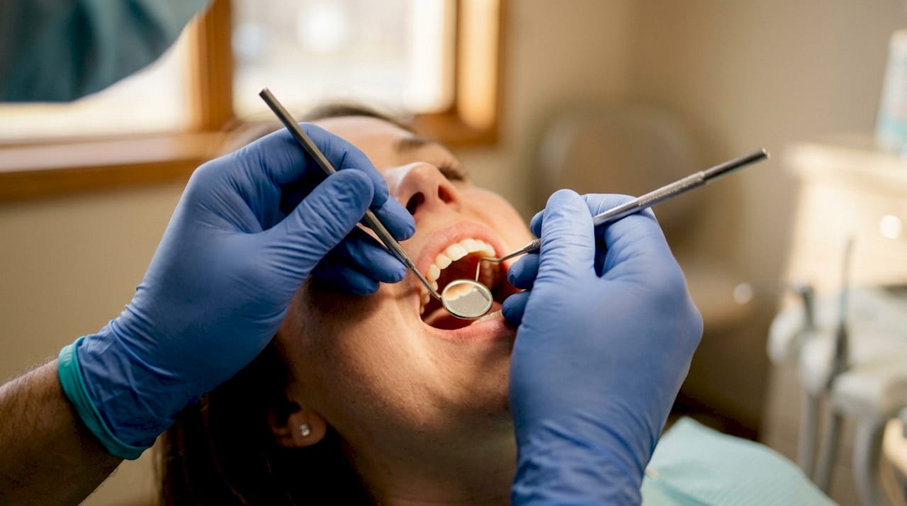
Gum health gets equal attention. Your dentist looks at the color, texture, and contour of your gum tissue. Healthy gums are firm and pink. Swollen, red, or bleeding gums signal inflammation, which can indicate gingivitis or early periodontal disease.
The most precise part of this assessment is periodontal probing. A thin, calibrated instrument called a periodontal probe is gently inserted between each tooth and the surrounding gum. Periodontal probing uses a 6-site method per tooth, recording pocket depths in millimeters at six points around each tooth. Your dentist or hygienist reads these numbers aloud as they work. Healthy tissue measures 1–3mm; a reading of 4mm or higher may indicate gum disease and requires closer attention.
Tooth mobility is also assessed using the Miller index, a standard clinical scale that grades how much a tooth moves when pressure is applied. Furcation involvement, which refers to bone loss between the roots of multi-rooted teeth, is another key finding that guides gum disease diagnosis and treatment decisions.
- Pocket depths of 1–3mm: healthy range
- Pocket depths of 4–5mm: early to moderate disease
- Pocket depths of 6mm or more: advanced periodontal disease
- Miller index grades tooth mobility from 0 (no movement) to 3 (severe)
Pro Tip: If you hear numbers being called out during your probing, ask your dentist to explain what they mean. A quick explanation turns those numbers into a clear picture of your gum health.
What does an oral cancer screening involve?
Oral cancer screening is a life-saving procedure that takes only 3–5 minutes during a routine exam. Many patients do not realize it is happening because it is woven into the soft tissue examination. The dentist systematically inspects and feels the tissues inside your mouth, looking for anything that does not belong.
The areas checked during an oral cancer screen include:
- The tongue, including the sides and underside
- The floor of the mouth
- The roof of the mouth (hard and soft palate)
- The inner cheeks and lips
- The back of the throat and tonsil area
Your dentist looks for red or white patches, lumps, sores, or any tissue that feels different from surrounding areas. Any suspicious lesion lasting more than two weeks without a clear cause typically leads to a biopsy referral. Early detection is the single most important factor in successful oral cancer treatment. This is one of the most compelling reasons to keep up with regular exams, even when your teeth feel fine.
Why do dentists take X-rays?
X-rays reveal what no visual exam can. Cavities between teeth, bone loss beneath the gum line, infections at the root tip, and impacted wisdom teeth are all invisible to the naked eye. Dental imaging fills that gap and often changes the treatment plan entirely.
The two most common types of dental X-rays are bitewing X-rays, which show the upper and lower back teeth and detect decay between teeth, and panoramic X-rays, which capture the full jaw, all teeth, and surrounding bone in a single image. Each type serves a different diagnostic purpose, and your dentist selects the right one based on what they need to see.
X-rays are not prescribed routinely but based on clinical needs and patient risk factors, following principles that minimize radiation exposure while maximizing diagnostic benefit. A child with no history of cavities and low risk factors may not need X-rays at every visit. An adult with a history of gum disease or multiple restorations may need them more frequently. The decision is always individualized.
- Bitewing X-rays: detect cavities between teeth and early bone loss
- Panoramic X-rays: assess jaw structure, all teeth, and impacted teeth
- Periapical X-rays: show the full length of individual teeth and surrounding bone
Pro Tip: If you are concerned about radiation, ask your dentist about digital X-rays. Digital systems use significantly less radiation than traditional film and produce clearer images.
How do dentists assess bite, jaw function, and facial structure?
The extraoral and bite assessment is the part of the exam most patients overlook. Dental exams extend beyond cavities to assess the entire oral-systemic health connection, including jaw joints and bite function, offering early detection of broader health issues. Your dentist checks how your upper and lower teeth fit together, a relationship called occlusion. A misaligned bite causes uneven wear, tooth fractures, and jaw pain over time.
Signs of teeth grinding (bruxism) show up clearly during this part of the exam. Worn, flattened tooth surfaces, cracked enamel, and sensitive teeth are all common findings. Identifying bruxism early allows your dentist to recommend a night guard before serious damage occurs.
The temporomandibular joint (TMJ) connects your jaw to your skull. Your dentist checks for clicking, popping, or limited movement when you open and close your mouth. The extraoral exam also includes palpation of lymph nodes and evaluation of facial symmetry, which can signal underlying conditions that extend well beyond the mouth.
| Exam component | What the dentist checks | Why it matters |
|---|---|---|
| Occlusion | How upper and lower teeth meet | Prevents uneven wear and fractures |
| TMJ assessment | Joint movement, clicking, pain | Detects jaw disorders early |
| Bruxism signs | Worn or cracked enamel | Guides night guard recommendations |
| Lymph node palpation | Swelling or tenderness in neck | Flags possible infection or pathology |
| Facial symmetry | Asymmetry in jaw or face | Identifies structural concerns |
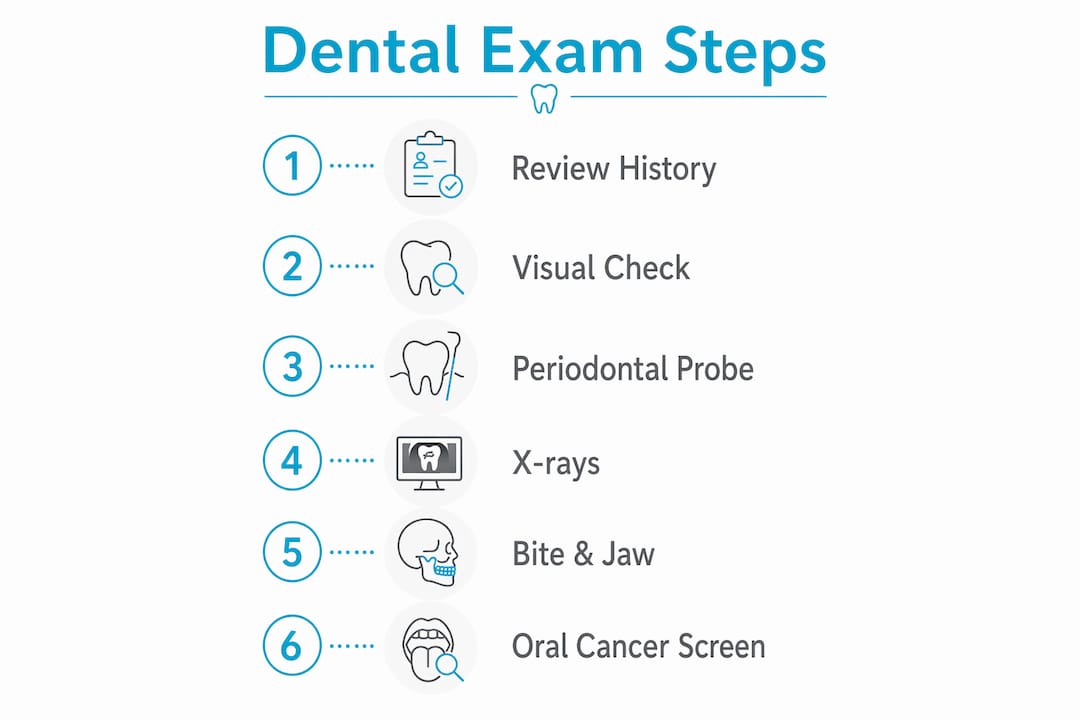
You can learn more about preparing for your dental visit to make the most of every component of your exam.
Key Takeaways
A thorough dental exam covers teeth, gums, soft tissues, bite, jaw function, and selective X-rays to detect problems early and guide personalized treatment for every patient.
| Point | Details |
|---|---|
| Exam scope is broad | Dentists assess teeth, gums, bite, jaw, soft tissues, and oral cancer risk in one visit. |
| Probing depths matter | Healthy gum pockets measure 1–3mm; readings of 4mm or higher signal potential disease. |
| Oral cancer screening is included | A 3–5 minute soft tissue check happens at every exam and can detect cancer early. |
| X-rays are individualized | Dentists order imaging based on your specific risk factors, not a fixed schedule. |
| History shapes the exam | Updated medical and dental history directly influences what your dentist looks for. |
What patients often miss about dental exams
In my experience, the biggest misconception patients bring into the exam room is that a checkup is just a cavity hunt. I have had patients sit down, open wide, and then look genuinely surprised when I start checking their neck, asking about jaw pain, or explaining what those probing numbers mean. That surprise tells me we have not done a good enough job explaining what we actually do.
The oral cancer screen is the one that concerns me most when patients skip appointments. Three to five minutes of examination can catch something that, found early, is highly treatable. Found late, the outcome changes dramatically. I have seen this firsthand, and it reinforces why I never rush that part of the exam.
The bite and TMJ assessment is another area where early findings prevent years of problems. A patient who grinds at night may not feel it. But the worn enamel tells the story clearly. Catching that at 35 is very different from catching it at 55 when the damage is extensive.
For families in Woodbridge and Vaughan, I always encourage bringing children in early and often. Kids’ exams follow the same systematic approach, scaled to their age and development. The habits and findings we document in childhood shape the care plan for decades. Regular exams are not just maintenance. They are the foundation of long-term oral health for the whole family.
You can read more about common dental issues families face and how early detection through exams makes a real difference.
— Felix
Comprehensive dental care for Woodbridge families
At Woodbridgedentalcentre, Dr. Michael Rouhi, Dr. Sandra Farber, and our team provide thorough dental exams for patients of all ages across Woodbridge, Vaughan, and Maple. Whether you are due for a periodic checkup or need a full comprehensive dental exam as a new patient, we take the time to explain every step and answer your questions.

Our clinic offers flexible appointment times and a welcoming environment designed to put both children and adults at ease. We believe every patient deserves to understand exactly what we find and why it matters. If your family is overdue for an exam, or if you want to know what to expect at your next visit, we are here to help. Reach out to Woodbridgedentalcentre to schedule your appointment and take a clear, confident step toward better oral health for everyone in your household.
FAQ
What does a standard dental exam include?
A standard dental exam includes a review of your medical history, visual inspection of teeth and gums, periodontal probing, oral cancer screening, bite and jaw assessment, and X-rays when clinically needed. Standard exams last 30–60 minutes, while new patient exams typically run 45–75 minutes.
How often should you get a dental exam?
Most patients benefit from a dental exam every six months, though your dentist may recommend more frequent visits based on your risk factors, gum health, or history of dental problems. The right interval depends on your individual clinical needs.
What is the difference between a comprehensive and a periodic exam?
A comprehensive exam establishes a full baseline with detailed charting, complete periodontal probing, oral pathology screening, and radiographic imaging. A periodic exam monitors your existing oral health status at regular intervals and is shorter in scope.
Do dentists always take X-rays at every exam?
No. X-rays are ordered based on clinical need and patient risk factors, not on a fixed schedule. Patients with low risk and no symptoms may go longer between imaging appointments than those with active dental concerns.
What questions should you ask your dentist after an exam?
Ask your dentist to explain your probing depths, any findings from the oral cancer screen, whether X-rays are needed, and what your biggest risk factors are. You can also find a helpful list of signs you may need a checkup to guide your conversation before the visit.


